|
|
_1.jpg) | Laboratory 9. Physics of Polymers and Polymeric Materials |
| Head of laboratory: Dr. Mariana CRISTEA |
|
|
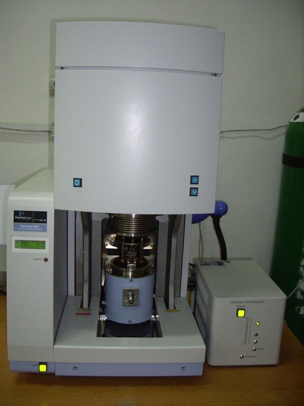
| Equipment: Dynamic mechanical analysis (DMA) |
|
|
- Dr. Mariana Cristea / mcristea@icmpp.ro |
|
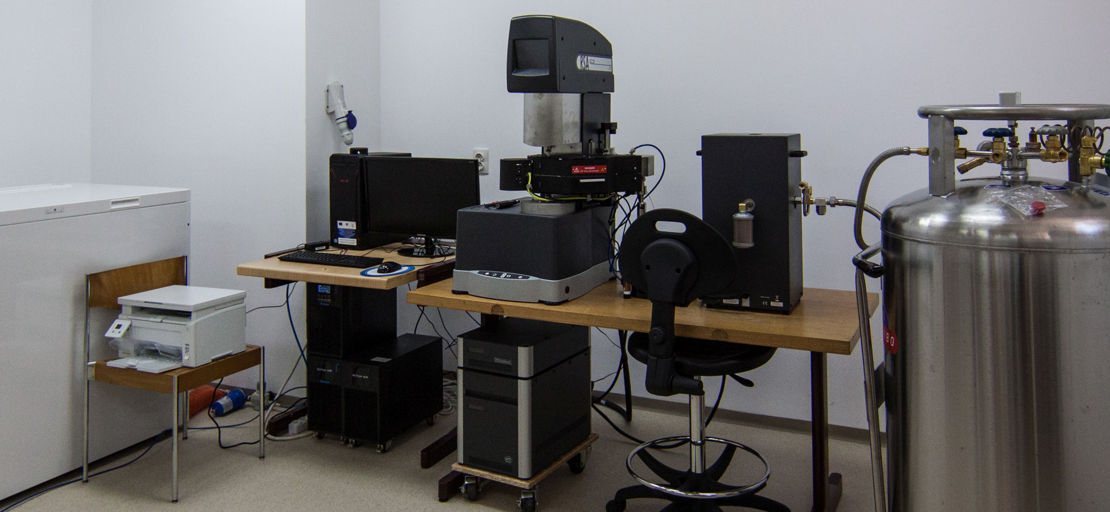
| Equipment: Rheometer for solid samples, RSA-G2 |
|
- Technique for the investigation of the viscoelastic properties of solid materials, especially polymeric materials;
- Temperature range: -150 oC ÷ 500 oC;
- Oven with camera viewer for image capture;
- Loading type: tension, shear, bending, compression;
- Information: evaluation of polymer relaxations and of activation energies, variation of storage modulus (E’) as a function of temperature, glass transition temperature determination, estimation of polymer properties in time.
|
- Dr. Mariana Cristea / mcristea@icmpp.ro |
|
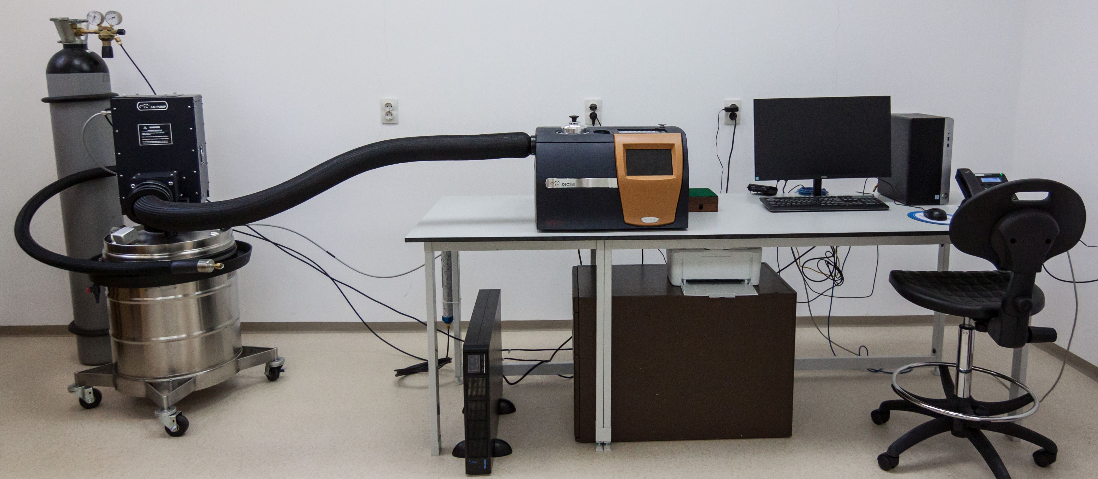
| Equipment: Differential Scanning Calorimeter, Discovery DSC 250 |
|
- Investigation of thermal behavior of polymers, organic and inorganic compounds, in terms of endothermic/exothermic processes, during isothermal regime and ramp/dynamic variation of temperature;
- Temperature range: -150 oC ÷ 400 oC, but not higher than the onset degradation temperature of the sample;
- Information: evaluation of the glass transition temperature, melting and crystallization processes, cure reactions and heats of transitions.
|
- Dr. Mariana Cristea / mcristea@icmpp.ro |
|
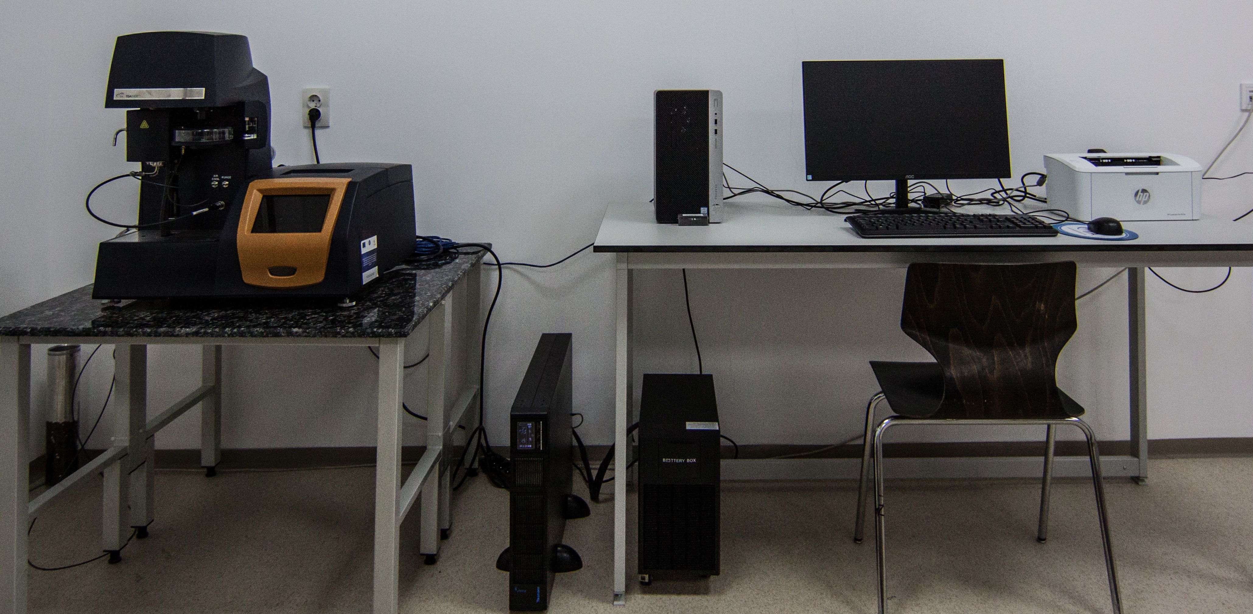
| Equipment: Thermogravimetric Analyzer, Discovery TGA 5500 |
|
- The instrument monitors the stability of a sample (polymers, organic and inorganic compounds) in terms of weight variation vs. time/temperature, in a controlled atmosphere;
- Temperature range: room temperature ÷ 1000 oC;
- Information: evaluation of the degradation temperatures, steps of degradation, determination of weight loss at a certain temperature, under specific conditions.
|
- Dr. Mariana Cristea / mcristea@icmpp.ro |
|
| Equipment: Agilent 6520 Accurate-Mass Q-TOF LC/MS |
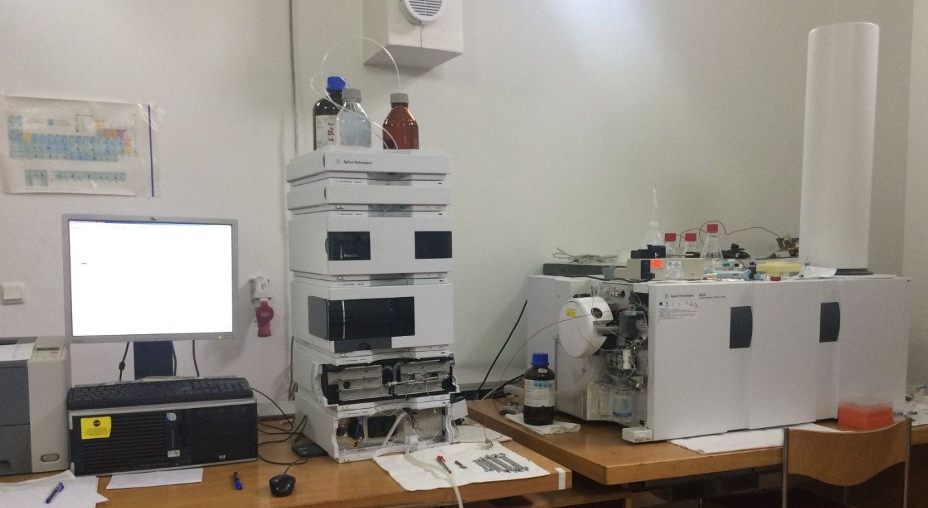
|
- Separation, identification and quantification of small organic/inorganic molecules and biomolecules;
- Electrospray Ionization (ESI);
- Hybrid quadrupole – Time of Flight (Q-TOF) analyzer;
- Resolution: up to 20,000 mass resolution;
- High sensitivity: up to 10-18 mol (scan mode);
- Mass accuracy: greater than 2 ppm;
- Mass range: up to 20,000 Da;
- Fast data acquisition (greater than or equal to 10 MS/MS spectra/sec) compatible with liquid chromatography (LC);
- Separation, identification and quantification of small organic/inorganic molecules and biomolecules;
- Electrospray Ionization (ESI);
- Hybrid quadrupole – Time of Flight (Q-TOF) analyzer;
- Resolution: up to 20000 mass resolution;
- High sensitivity: up to 10-18 mol (scan mode);
- Mass accuracy: greater than 2 ppm;
- Mass range: up to 20,000 Da;
- Fast data acquisition (greater than or equal to 10 MS/MS spectra/sec) compatible with liquid chromatography (LC).
|
- Dr. Mihaela Silion / silion.mihaela@icmpp.ro |
|
| Equipment: Verios G4 UC Scanning Electron Microscope (Thermo Fisher Scientific) equipped with an energy dispersive spectrometer (EDS, EDAX Octane Elite) |
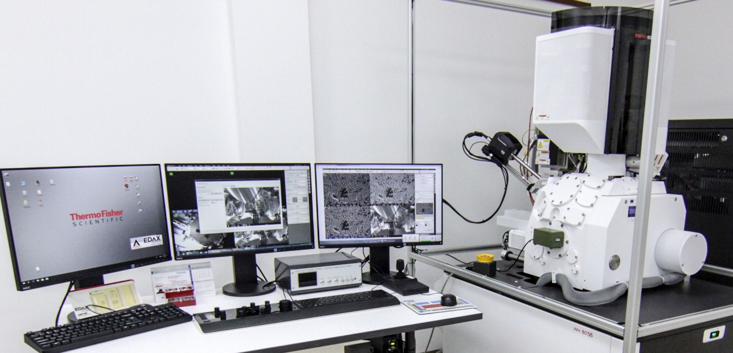
|
The microscope allows the complete, high resolution morphological investigations of a broad variety of samples. It also affords information regarding samples’ composition based on several BF/DF/HAADF-STEM and EDX detectors.
Specifications:
• Electron source: Schottky thermal field emitter;
• Accelerating voltage range: 0.2 to 30 kV;
• Detectors:
- Elstar in-lens SE detector (TLD-SE);
- Elstar in-lens BSE detector (TLD-BSE);
- Elstar in-column SE detector (ICD);
- Elstar in-column BSE detector (MD);
- Everhart-Thornley SE detector (ETD);
- Retractable, low voltage, high-contrast, solid-state backscatter electron detector (DBS);
- Retractable STEM detector with BF / DF / HAADF segments;
- IR camera for viewing sample/column;
- Chamber mounted Nav-Cam+™ .
• Resolution: 0.6 nm at 15 kV. |
- Dr. Florica Doroftei / florica.doroftei@icmpp.ro |
|
| Equipment: Quanta 200 Scanning Electron Microscope (FEI Company) equipped with an energy dispersive spectrometer (EDS, EDAX silicon drift detector) |
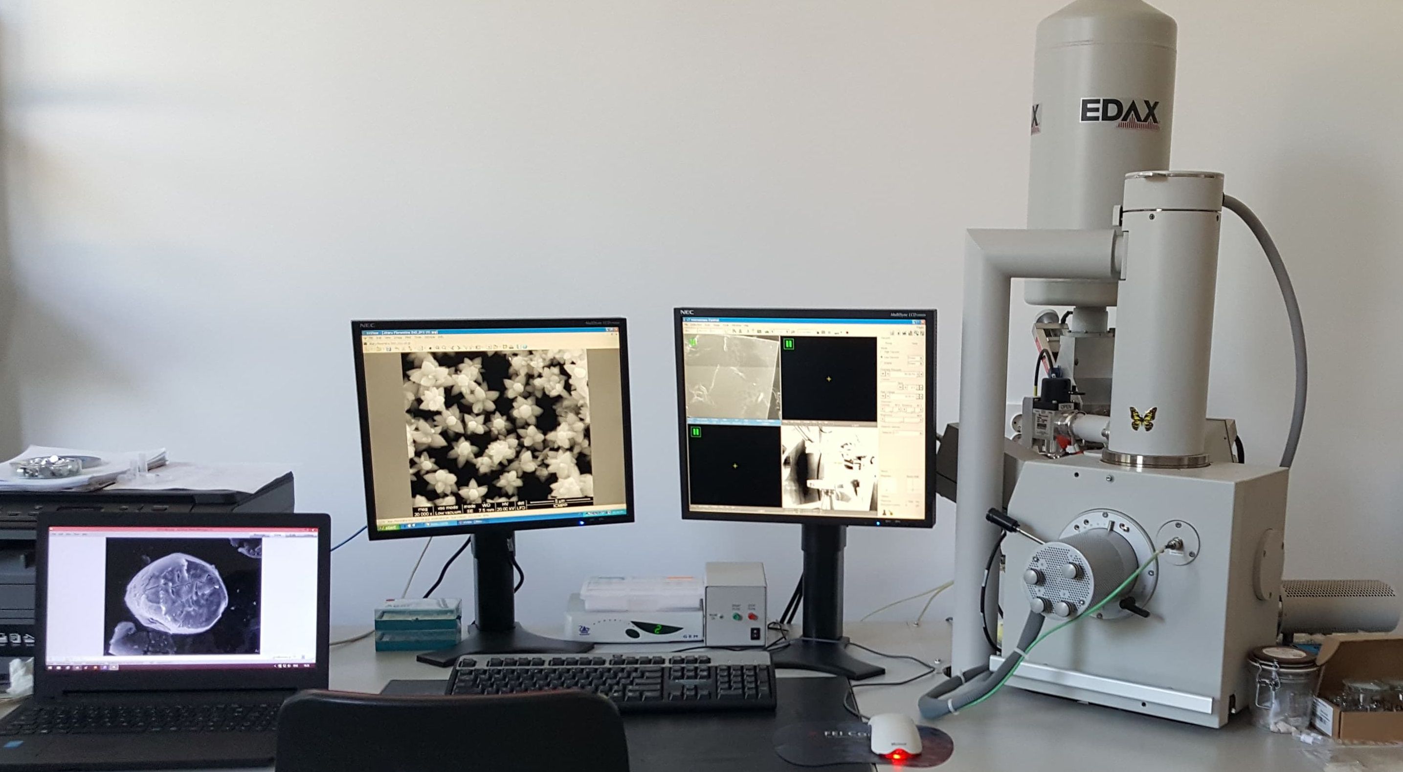
|
The microscope enables morphological investigations and X-ray microanalysis for a wide range of samples and can be operated in three vacuum modes: high vacuum (HV), low vacuum (LV) and environmental (ESEM). Energy-dispersive X-ray analysis (EDX) may be undertaken at both high and low vacuum to determine elemental compositions and compositional maps.
Specifications:
• Electron source: Tungsten filament;
• Accelerating voltage range: 0.2 to 30 kV;
• Pressure in low vacuum mode: <200 Pa;
• Detectors:
- Everhart Thornley Detector (ETD): secondary electrons (SE), backscattered electrons (BSE) in HV;
- Large Field Detector (LFD): SE in LV;
- Solid State Backscattered Electron Detector (BSED): BSE in HV and LV;
- Gaseous Secondary Electron Detector (GSED): SE in ESEM;
- EDS Detector for EDAX measurements;
• Resolution: 4nm at 30 kV. |
- Dr. Florica-Mirela Doroftei / florica.doroftei@icmpp.ro |
|
| Equipment: Leica EM UC7 Ultramicrotome with a Leica EM FC7 cryo chamber for room temperature and cryo sectioning |
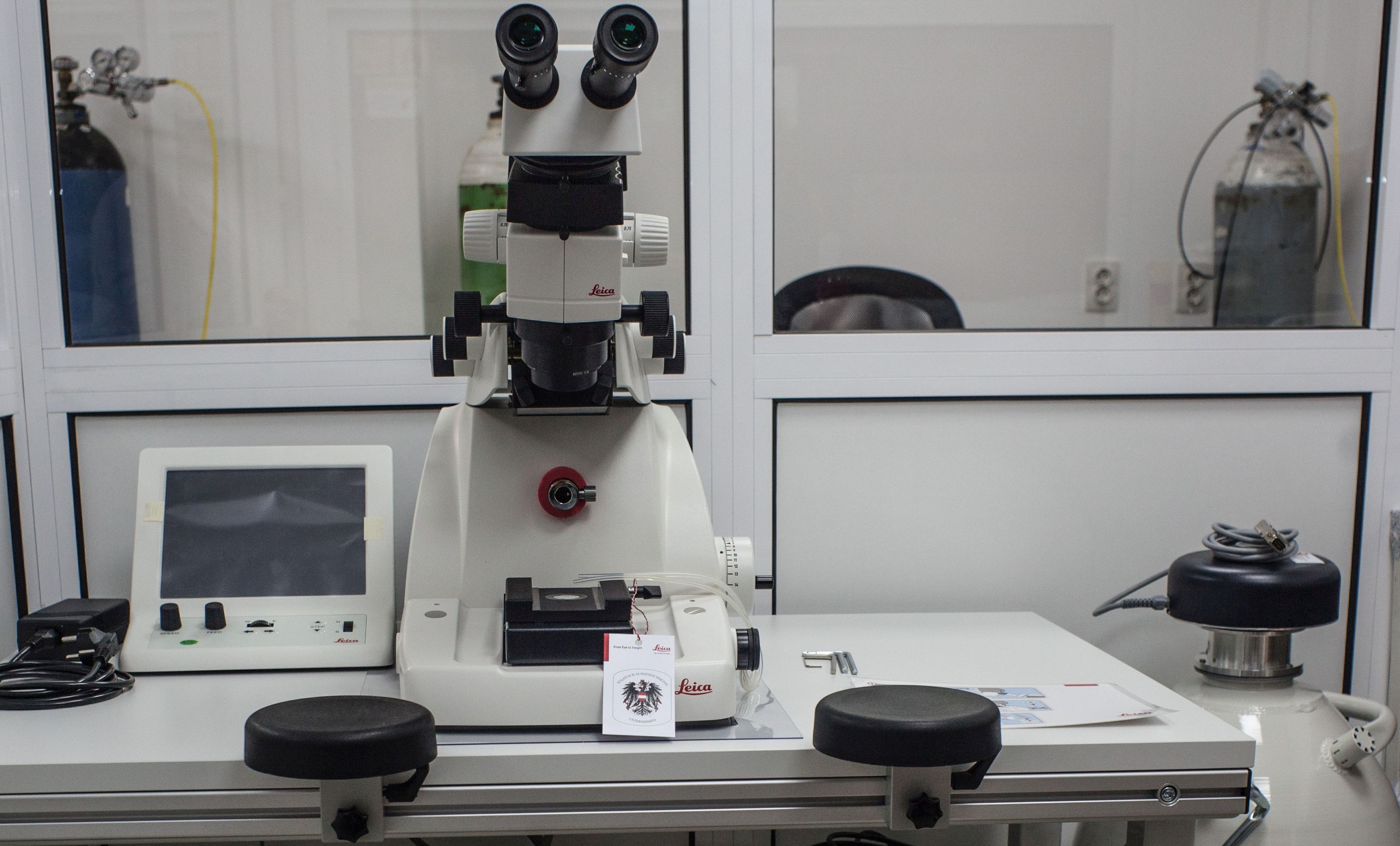
|
The device permits the preparation of semi- and ultra-thin sections in materials as required by TEM, SEM and AFM techniques. |
- Dr. Florica Doroftei / florica.doroftei@icmpp.ro |
|
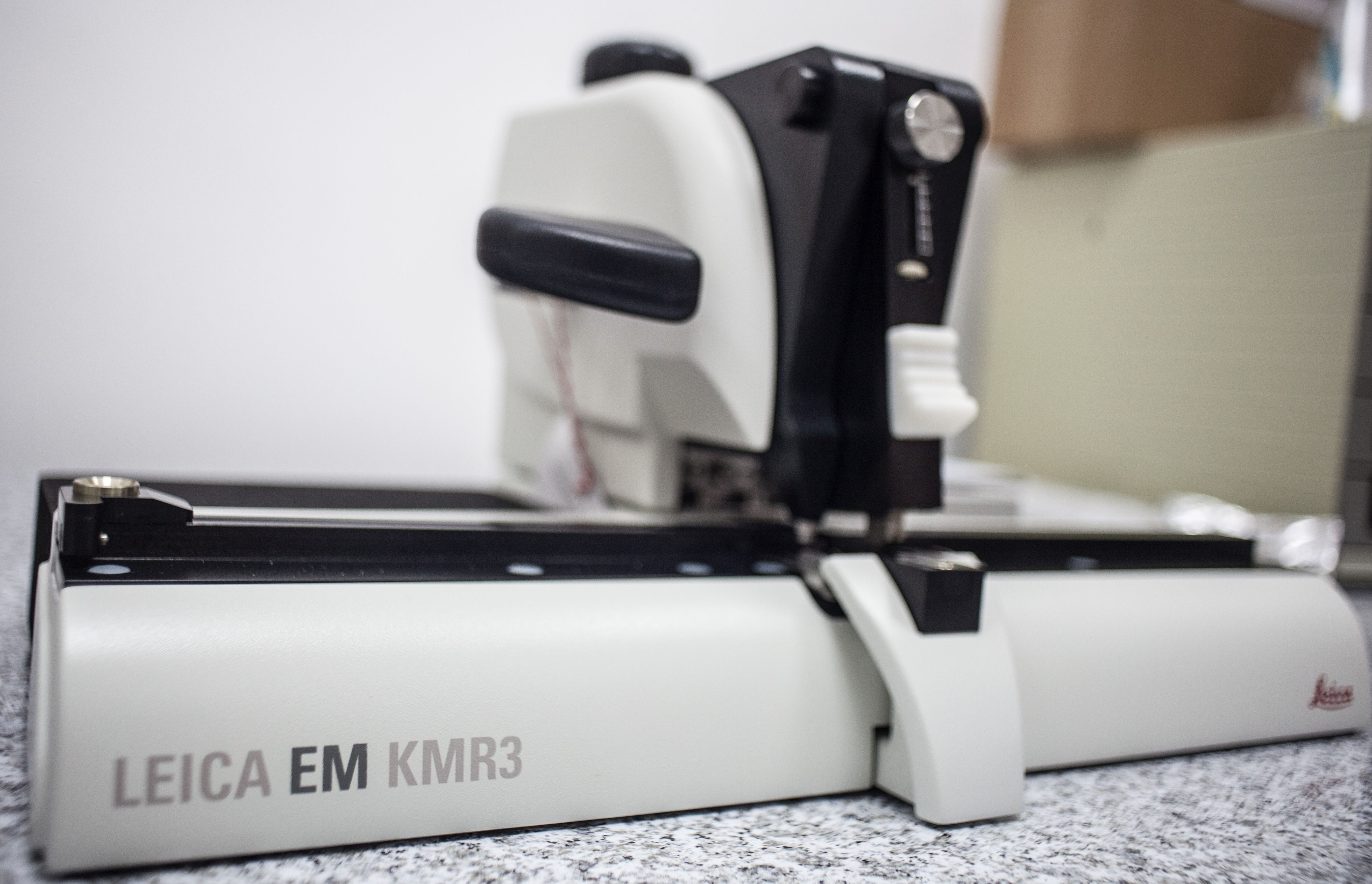
| Equipment: Leica EM KMR3 Glass Knife Maker |
|
The device used to make glass knives for ultramicrotome in two thicknesses: 6.4 mm and 8 mm. |
- Dr. Florica Doroftei / florica.doroftei@icmpp.ro |
|
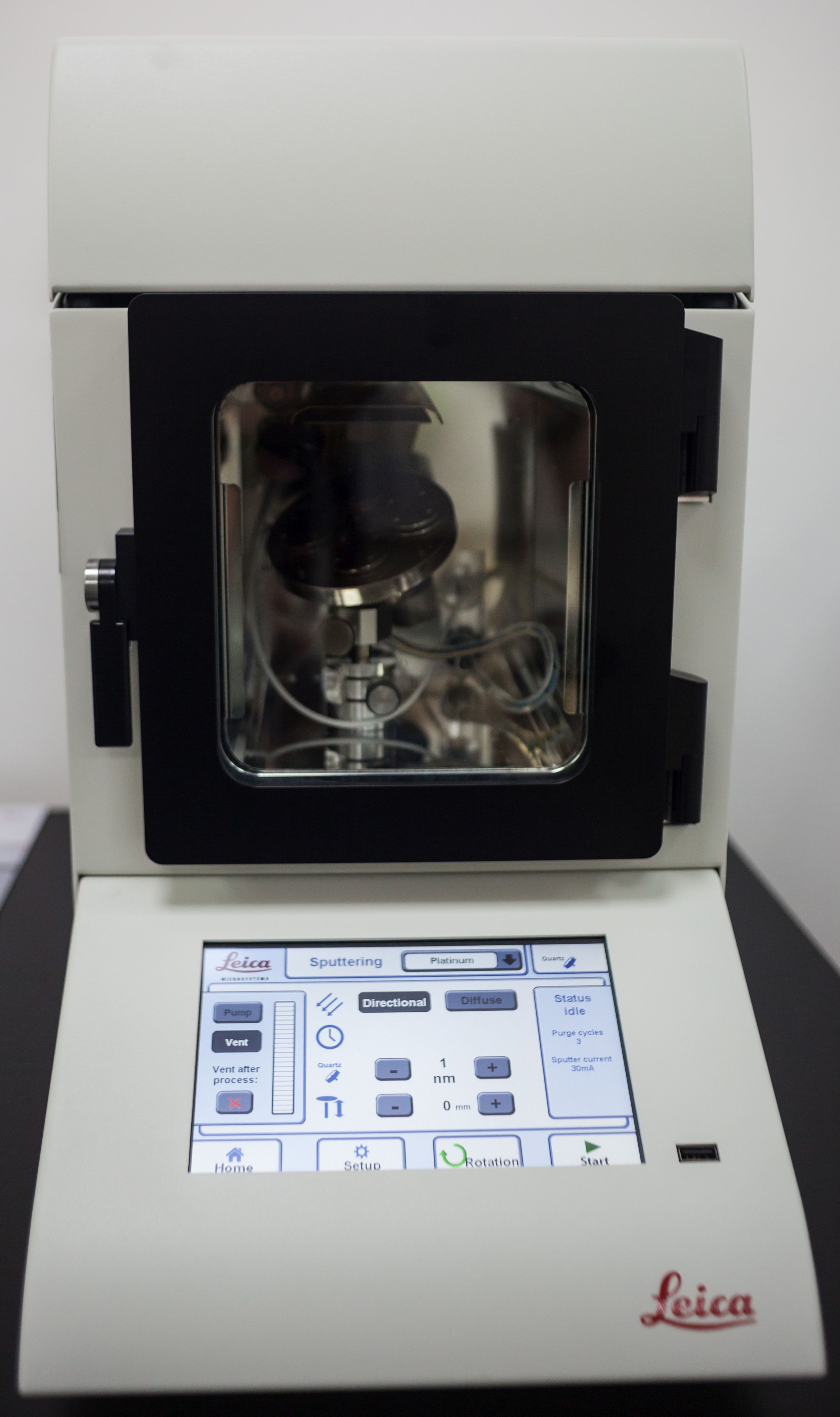
| Equipment: Leica EM ACE200 Sputter Coater |
|
The coater is designed to produce homogenous coatings of conductive metals (Au, Pt, Pd) or carbon for SEM and TEM analysis. It can be configured as a sputter coater or a carbon thread evaporation coater. |
- Dr. Florica Doroftei / florica.doroftei@icmpp.ro |
|
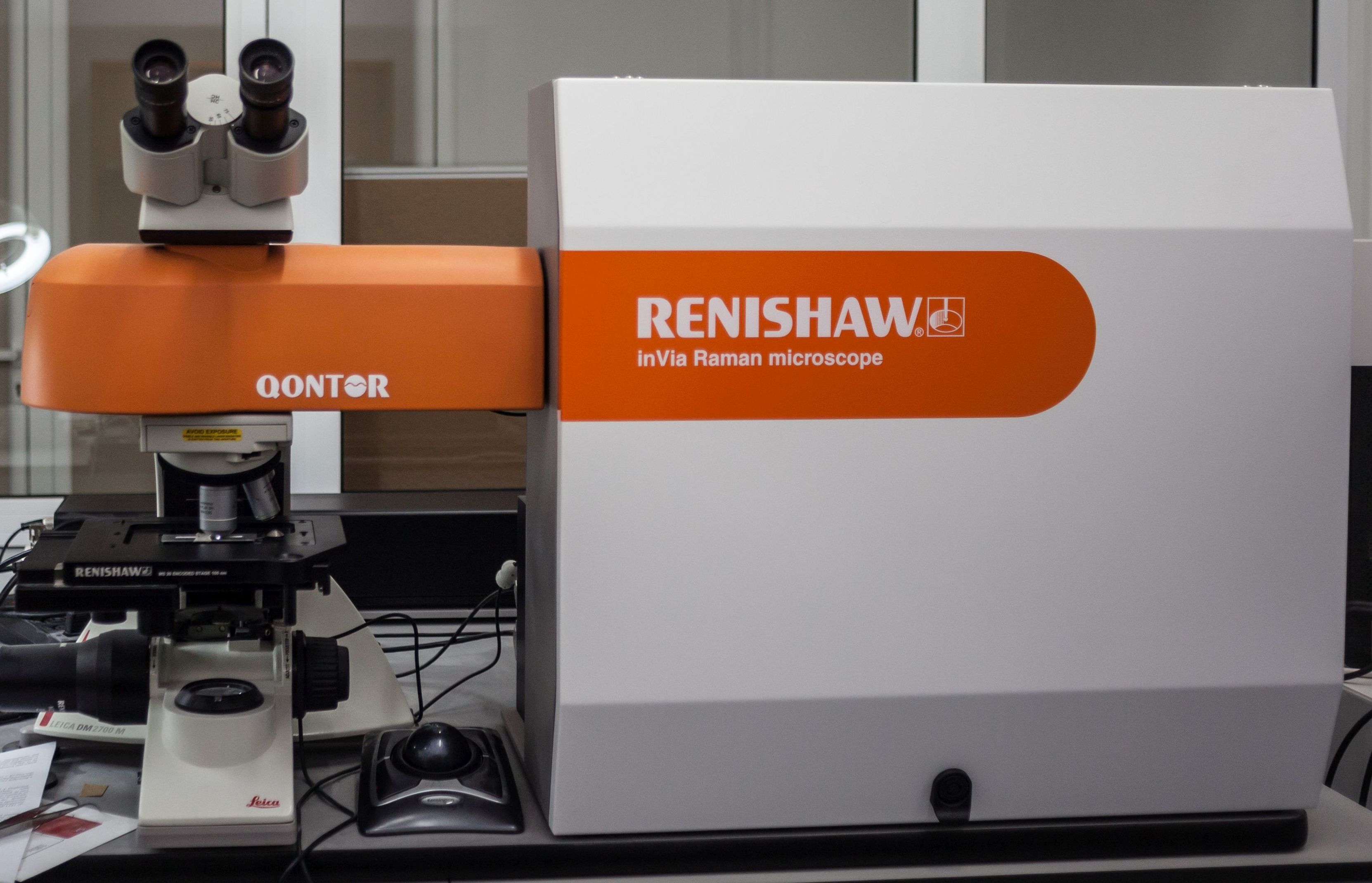
| Equipment: Raman spectrometer with confocal microscope with ultra-fast autofocus and optical profilometry Renishaw inVia Qontor |
|
- 633 nm He-Ne laser, 17 mW;
- Spectral range: 100 ÷ 5000 cm-1;
- Leica confocal microscope with a resolution below 2 µm;
- Stigmatic Raman spectrometer, with real-time autofocus and 3D surface extraction mode;
- Renishaw Wire 5.0 software for control and acquisition;
- Soft Stream HR;
- The system performs both spectroscopic imaging and imaging for the 3D profile of the surface.
|
- Dr. Magdalena Aflori / maflori@icmpp.ro |
|
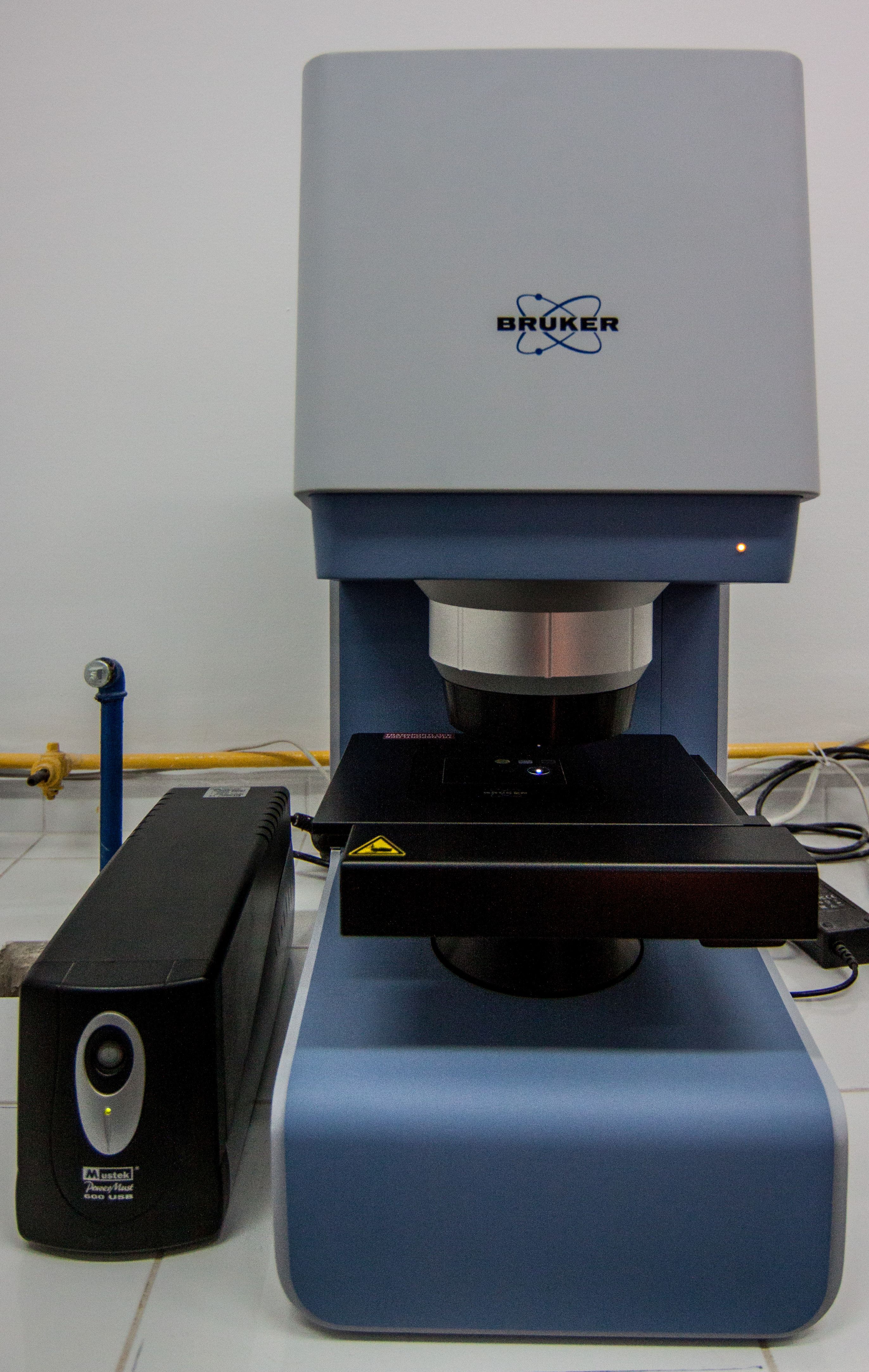
| Equipment: Bruker Lumos FTIR spectrometer microscope |
|
- Spectral investigation in transmission, reflection and ATR of multilayer materials, films, membranes, to determine several components in an inhomogeneous material, the distribution of components in a biological tissue, identification of unknown material;
- Spectral range: 7800 ÷ 450 cm-1;
- Wave number accuracy: 0.05 cm-1;
- Spectral resolution: 2 cm-1.
|
- Dr. Magdalena Aflori / maflori@icmpp.ro |
|
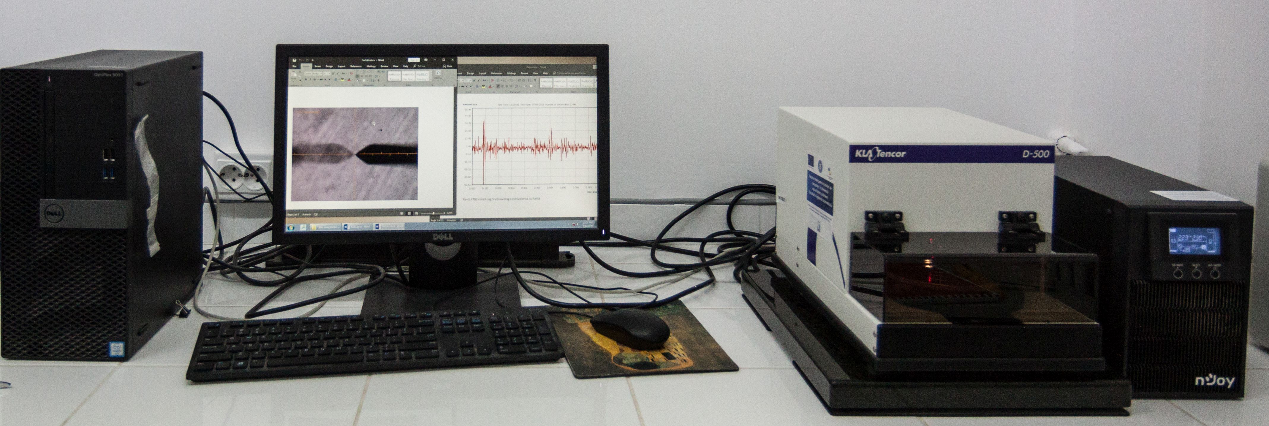
| Equipment: KLA Tencor D500 contact profiler |
|
- Vertical scanning range up to 1200 μm;
- Scan length: up to 30 mm;
- Side optical view of the sample with wide field of view (3760 x 3120 μm);
- 5 MP color digital video camera with 4x digital zoom and lighting control;
- Determines the thickness of polymer films, thin layers; characterizes the topography of thin deposits by evaluating the profile, roughness, surface texture, on hard samples, as well as on soft samples, by 2D measurements of surface profile parameters at nanometric level, in steps, in a range between nanometers and micrometers; allows the evaluation of surface uniformity and stress in thin films.
|
- Dr. Magdalena Aflori / maflori@icmpp.ro |
|
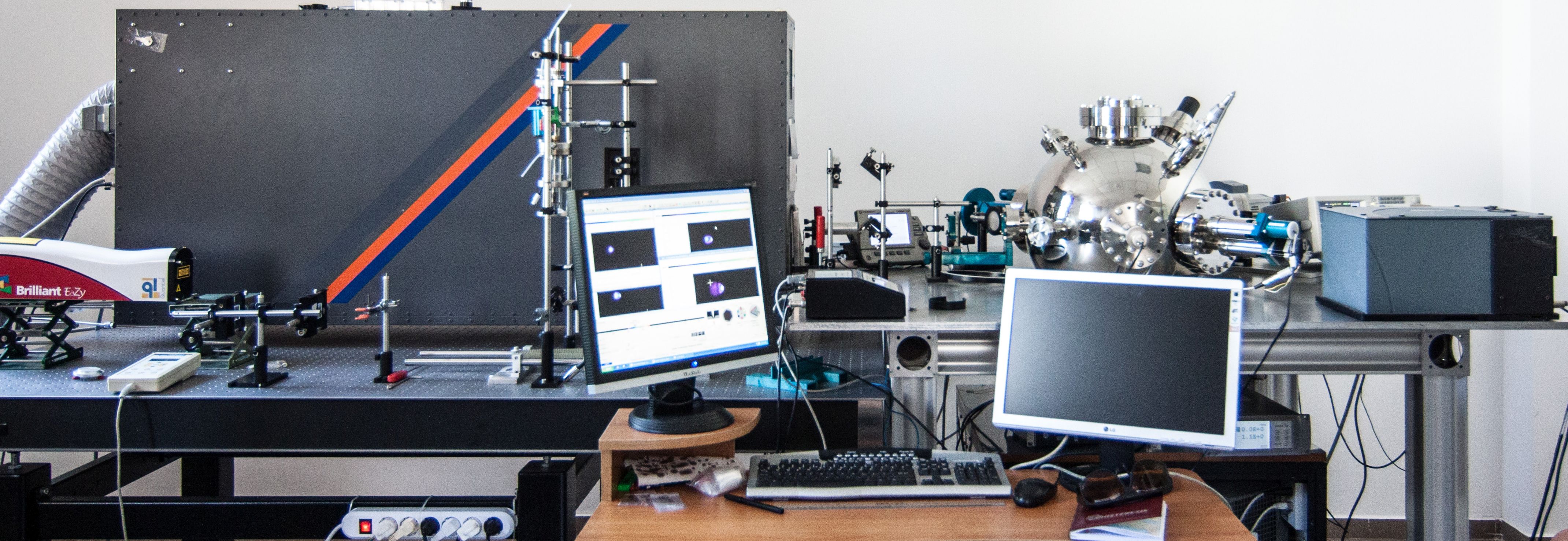
| Equipment: Excimers lasers laboratory |
|
- System for thin film deposition through laser ablation;
- VarioLas laser lithography system;
- System for generation of nanoparticles in suspension through liquid laser ablation.
|
- Dr. Mihaela-Adriana Olaru / olaruma@icmpp.ro
- Dr. Cristian Ursu / cristian.ursu@icmpp.ro |
|
|